The System:
All of the structures involved in moving air into and out of the lungs, and the exchange of gases between cells. It provides large surface area for gas exchange, moves air to and from gas exchange surfaces, defends and protects gas exchange surfaces from dehydration, temperature changes, and pathogens. It aids in providing the sounds necessary for speech and other forms of communication as well as aiding the sense of smell in the nasal cavity.
Major Structures include:
Nose, Pharynx, Larynx, Trachea, Bronchi, Bronchioles, Lungs, Alveoli
The respiratory tract includes all of the passageways that carry air between the nose/mouth and the lungs. The conducting portion includes the nasal cavity to the bronchi, and the respiratory portion to the bronchioles and alveoli.
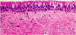
The respiratory mucosa lines the conducting portion and consists of ciliated columnar epithelium and goblet cells. The mucus traps debris and pathogens in the nasal cavity and the cilia sweeps the mucus into the pharynx where it is destroyed by being expelled or swallowed.
All of the structures involved in moving air into and out of the lungs, and the exchange of gases between cells. It provides large surface area for gas exchange, moves air to and from gas exchange surfaces, defends and protects gas exchange surfaces from dehydration, temperature changes, and pathogens. It aids in providing the sounds necessary for speech and other forms of communication as well as aiding the sense of smell in the nasal cavity.
Major Structures include:
Nose, Pharynx, Larynx, Trachea, Bronchi, Bronchioles, Lungs, Alveoli
The respiratory tract includes all of the passageways that carry air between the nose/mouth and the lungs. The conducting portion includes the nasal cavity to the bronchi, and the respiratory portion to the bronchioles and alveoli.
The respiratory mucosa lines the conducting portion and consists of ciliated columnar epithelium and goblet cells. The mucus traps debris and pathogens in the nasal cavity and the cilia sweeps the mucus into the pharynx where it is destroyed by being expelled or swallowed.
The Nose
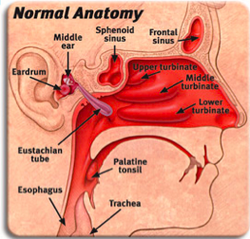
The external nares are the nostrils. The nasal vestibule is a flexible tissue where nose hairs protect against large particles and debris and the nasal cavity is the space where air is warmed and filtered before entering the respiratory tract. The frontal sinuses are mucus producing cavities in the front of the skull that reduce the weight of the skull and can also amplify sounds through resonance
The external nares are the nostrils. The nasal vestibule is a flexible tissue where nose hairs protect against large particles and debris and the nasal cavity is the space where air is warmed and filtered before entering the respiratory tract. The frontal sinuses are mucus producing cavities in the front of the skull that reduce the weight of the skull and can also amplify sounds through resonance
- The nose is the only part of the respiratory system that you can see. It is where air enters but it is also where air is warmed, made moist, filtered and cleaned, and smelled.
- The nose has tiny hairs in it that clean the air as you breathe in. This prevents some of the intrusion of dust and other particles from getting into the body/lungs. It is impossible to prevent any harmful pathogens entering , but the nose is a great aid defense for the body when breathing.
- Air can be inhaled or exhaled here, and the air exiting the body through the nose returns moisture and heat to the nasal cavity before being exhaled into the environment.
- The nose is a structure of the face made of cartilage, bone, muscle, and skin that supports and protects the anterior portion of the nasal cavity. The nasal cavity is a hollow space within the nose and skull that is lined with hairs and mucus membrane.
The Pharynx
The pharynx is just a passageway for air to pass through, but it is ringed by tonsils that trap and kill bacteria that come in with the air. It is apart of both the digestive and respiratory system.
The pharynx is just a passageway for air to pass through, but it is ringed by tonsils that trap and kill bacteria that come in with the air. It is apart of both the digestive and respiratory system.
- It is divided into three sections: The nasopharynx
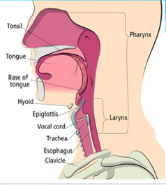
occupies the posterior end of the nasal cavity, the oropharynx extends from the oral cavity to the hyoid bone, and the laryngopharynx extends from hyoid to the opening of the esophagus
- It is also known as the throat, and is a muscular funnel that extends from the posterior end of the nasal cavity to the superior end of the esophagus and larynx.
- It is then diverted into the opening of the larynx by the epiglottis. The epiglottis is a flap of elastic cartilage that acts as a switch between the trachea and the esophagus. Because the pharynx is also used to swallow food, the epiglottis ensures that air passes into the trachea by covering the opening to the esophagus. During the process of swallowing, the epiglottis moves to cover the trachea to ensure that food enters the esophagus and to prevent choking.
The LarynxThe larynx is a doorway. When food is passing through the pharynx, the larynx closes. When we inhale air, it is open and passes through toward the lungs. The larynx also has the vocal cords that make sound for speech. After air passes through the larynx, they enter the trachea. The epiglottis is the protective flap that covers the opening to the larynx when swallowing food or
drink. The glottis is the narrow opening into the larynx. The false vocal chords
are inelastic ligaments that protect the glottis from foreign objects, and the true vocal chords are elastic
ligaments whose tension could be altered by muscles. Air causes vibration, vibration produces
sound.
- Also known as the voice box, is a short section of the airway that connects the laryngopharynx and the trachea.
- The larynx is located in the anterior portion of the neck, just inferior to the hyoid bone and superior to the trachea. Those several cartilage structures above make up the larynx and give it its structure.
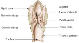
- Inferior to the epiglottis is the thyroid cartilage, which is often referred to as the Adam’s apple as it is most commonly enlarged and visible in adult males. The thyroid holds open the anterior end of the larynx and protects the vocal folds. Inferior to the thyroid cartilage is the ring-shaped cricoid cartilage which holds the larynx open and supports its posterior end. In addition to cartilage, the larynx contains special structures known as vocal folds, which allow the body to produce the sounds of speech and singing.
- The vocal folds are folds of mucous membrane that vibrate to produce vocal sounds. The tension and vibration speed of the vocal folds can be changed to change the pitch that they produce.
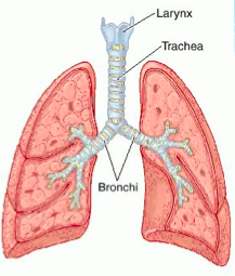
The Trachea
The trachea (windpipe) is about 4 inches long (12 cm) and is kept open by tiny rings of stiff cartilage. The last ring of cartilage is called the carina and is covered by sensitive tissue. If you inhale anything but air, when it touches the carina, you cough violently until it is gone. This protects the lower airways from being blocked. It is a tough, flexible tube of cartilage from the base of the larynx that branches into the two bronchi. Tracheal Cartilage is a C-shaped wall of cartilage that prevents the trachea from collapsing. The trachealis muscle is smooth muscle tissue that can adjust the diameter of the trachea, and regulates the amount of air passing through the respiratory tract.
The trachea splits at the bottom to become the two bronchi, one for each lung. The primary bronchi are large passageways that diverge at the base of the trachea, and have the same structure as the trachea. They branch into smaller airways called the bronchial tree. The secondary bronchi are smaller airways that begin at the entrance to the lungs, tertiary bronchi are even smaller airways at the center of the lungs. They also keep lungs suspended in chest cavity.
The trachea (windpipe) is about 4 inches long (12 cm) and is kept open by tiny rings of stiff cartilage. The last ring of cartilage is called the carina and is covered by sensitive tissue. If you inhale anything but air, when it touches the carina, you cough violently until it is gone. This protects the lower airways from being blocked. It is a tough, flexible tube of cartilage from the base of the larynx that branches into the two bronchi. Tracheal Cartilage is a C-shaped wall of cartilage that prevents the trachea from collapsing. The trachealis muscle is smooth muscle tissue that can adjust the diameter of the trachea, and regulates the amount of air passing through the respiratory tract.
- lined with pseudostratified ciliated columnar epithelium.
- The rings of cartilage making up the trachea allow it to remain open to air at all times. The open end of the cartilage rings faces posteriorly toward the esophagus, allowing the esophagus to expand into the space occupied by the trachea to accommodate masses of food moving through the esophagus.
- The main function of the trachea is to provide a clear airway for air to enter and exit the lungs. In addition, the epithelium lining the trachea produces mucus that traps dust and other contaminants and prevents it from reaching the lungs. Cilia on the surface of the epithelial cells move the mucus superiorly toward the pharynx where it can be swallowed and digested in the gastrointestinal tract.
The trachea splits at the bottom to become the two bronchi, one for each lung. The primary bronchi are large passageways that diverge at the base of the trachea, and have the same structure as the trachea. They branch into smaller airways called the bronchial tree. The secondary bronchi are smaller airways that begin at the entrance to the lungs, tertiary bronchi are even smaller airways at the center of the lungs. They also keep lungs suspended in chest cavity.
- At the inferior end of the trachea, the airway splits into left and right branches known as the primary bronchi.
- The left and right bronchi run into each lung before branching off into smaller secondary bronchi. The secondary bronchi carry air into the lobes of the lungs—2 in the left lung and 3 in the right lung. The secondary bronchi in turn split into many smaller tertiary bronchi within each lobe.
- The tertiary bronchi split into many smaller bronchioles that spread throughout the lungs. Each bronchiole further splits into many smaller branches less than a millimeter in diameter called terminal bronchioles. Finally, the millions of tiny terminal bronchioles conduct air to the alveoli of the lungs.
- As the bronchi branch into secondary and tertiary bronchi, the cartilage becomes more widely spaced and more smooth muscle and elastin protein is found in the walls.
The Bronchioles
The right and left bronchi enter the lungs and branch many times into smaller and smaller bronchioles before reaching the tiny ends where oxygen exchange happens. They are the smallest airways inside the lungs and supply air directly to the lung tissue. Lobule is a portion of lung tissue that is bound to a bronchiole by connective tissue. The terminal bronchiole connected to the lobule diverges into respiratory bronchioles.
The right and left bronchi enter the lungs and branch many times into smaller and smaller bronchioles before reaching the tiny ends where oxygen exchange happens. They are the smallest airways inside the lungs and supply air directly to the lung tissue. Lobule is a portion of lung tissue that is bound to a bronchiole by connective tissue. The terminal bronchiole connected to the lobule diverges into respiratory bronchioles.
- The bronchioles differ from the structure of the bronchi in that they do not contain any cartilage at all. The presence of smooth muscles and elastin allow the smaller bronchi and bronchioles to be more flexible and contractile.
- The main function of the bronchi and bronchioles is to carry air from the trachea into the lungs. Smooth muscle tissue in their walls helps to regulate airflow into the lungs. When greater volumes of air are required by the body, such as during exercise, the smooth muscle relaxes to dilate the bronchi and bronchioles. The dilated airway provides less resistance to airflow and allows more air to pass into and out of the lungs.
- The smooth muscle fibers are able to contract during rest to prevent hyperventilation. The bronchi and bronchioles also use the mucus and cilia of their epithelial lining to trap and move dust and other contaminants away from the lungs.
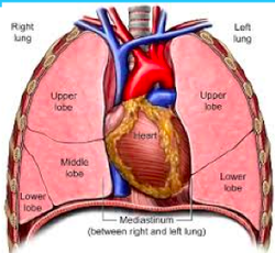
The Lungs
The lungs are a pair of large, spongy organs found in the thorax lateral to the heart and superior to the diaphragm. Each lung is surrounded by a pleural membrane that provides the lung with space to expand as well as a negative pressure space relative to the body’s exterior. The negative pressure allows the lungs to passively fill with air as they relax. The left and right lungs are slightly different in size and shape due to the heart pointing to the left side of the body. The left lung is therefore slightly smaller than the right lung and is made up of 2 lobes while the right lung has 3 lobes.
The lungs are a pair of large, spongy organs found in the thorax lateral to the heart and superior to the diaphragm. Each lung is surrounded by a pleural membrane that provides the lung with space to expand as well as a negative pressure space relative to the body’s exterior. The negative pressure allows the lungs to passively fill with air as they relax. The left and right lungs are slightly different in size and shape due to the heart pointing to the left side of the body. The left lung is therefore slightly smaller than the right lung and is made up of 2 lobes while the right lung has 3 lobes.
- Each lung is divided into sections called lobes. The right lung has 3 lobes. The left lung has 2.
- Rounded ends and indentations are caused by the shape of ribcage and other organs (heart, arteries)
- Spongy texture due to moisture required for gas exchange
- Significant amounts of elastic fibers allow for the increase of volume and surface area when required
- Parietal pleura – covers the inner surface of the thoracic cavity, diaphragm, and mediastinum
- Visceral pleura – covers the outside surface of the lungs
- The right and left pleural cavities are separated by the mediastinum
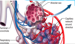
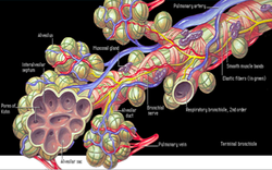
AlveoliThe interior of the lungs is made up of spongy tissues containing many capillaries and around 30 million tiny sacs known as alveoli. The alveoli are cup-shaped structures found at the end of the terminal bronchioles and surrounded by capillaries. The alveoli are lined with thin simple squamous epithelium that allows air entering the alveoli to exchange its gases with the blood passing through the capillaries.
- Respiratory bronchioles feed air into alveolar ducts. The ducts lead into alveolar sacs, which are chambers filled with several alveoli.
- Alveoli – cellular clusters that serve as the gas exchange surface in the lungs
- Alveolar macrophages – dust cells that patrol the alveoli, removing debris and pathogens
- Septal cells – large cells that secrete surfactant, which keeps alveoli moist and reduces surface tension
- Each lung has approx. 150 Million alveoli
- Each alveoli has a respiratory membrane with 3 layers:
*Thin simple squamous epithelium on the outside
*Endothelium lining the capillary on the inside
*Fused basement membrane between the first two layers - Designed for rapid diffusion of gases
Diaphragm
Is the smooth muscle that divides the thoracic and abdominal cavities. Controls the volume of the thoracic cavity and lungs. Dome shaped when relaxed, lungs are empty. Flattens when contracting, lungs expand as air flows in.
Rib Cage
It elevates and depresses to control volume in thoracic cavity. Synchronizing with the diaphragm maximizes air movement
Compliance – the ability of the lungs to expand. Determines athleticism or whether or not you have a disease
Tidal Volume – the amount of air moved in and out of the lungs in a single respiratory cycle (inhale/exhale)
Vital Capacity – Maximum amount of air that can be held in the lungs
Quiet breathing/Forced breathing – the difference between breathing passively or voluntarily. Determines the muscles involved and energy being used
Carbon dioxide is transported in blood 3 ways:
*Dissolved in plasma
*Binds to hemoglobin
*Converted to carbonic acid by erythrocytes
Respiratory centers in the brain are sensitive to concentration of CO2 in blood (pCO2)
High CO2 concentration will caused the brain to increase breathing. However, there are other factors that control this too (emotions, disease, etc.)
